4184547
Description
Flashcards by Sophie Burk, updated more than 1 year ago
|
|
Created by Sophie Burk
almost 9 years ago
|
|
| Question | Answer |
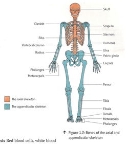
| Differentiate between the axial and appendicular skeleton | axial skeleton: protects vital organs and supports muscles appendicular skeleton: provides a framework for movement, supports muscles |
| Define long bones | longer than width, growth plates at both ends with hyaline cartilage to absorb shock and protect against friction humerus, femur, phalanges |
| Define short bones | as wide as long, provides support and stability with little movement, thin layer of compact bone tarsals, carpals |
| Define flat bones | strong plates of bone, in and around vital organs sternum, cranium, scapula, ribs |
| Define irregular bones | spongy/cancellous bone, thin outer layer of compact bone vertebrae, mandible, sacrum |
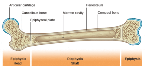
| (Bone Diagram) | |
| Describe articular cartilage | protects against friction, in synovial joints only |
| Define spongy/cancellous bone | many holes, osseous tissue, in the epiphysis |
| Describe the epiphysis of a bone | rounded end, works in ball + socket joints |
| Describe the diaphysis of a bone | main midsection of the bone, shaft, filled with bone marrow |
| Define bone marrow | produces WBC + RBC + platelets, flexible tissue |
| Define the periosteum | dense connective tissue that covers the bone |
| Define a compact bone | 80% of bones in the human body, made of calcium salts and inorganic substances, osseous tissue |
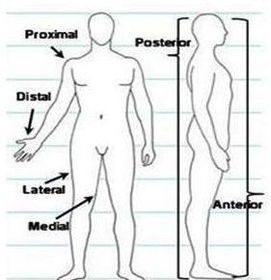
| (Anatomical Terminology of Bones Diagram) | |
| Define 'superior' | a structure above another, nearer to skull -cranium is superior to scapula |
| Define 'inferior' | structure below another, nearer to feet -tarsals inferior to carpals |
| Define 'proximal' | nearer to point of attachment of another structure -femur proximal to tibia |
| Define 'distal' | farther from point of attachment than another structure -metatarsal distal to humerus |
| Define 'medial' | nearer to midline than another structure -sternum medial to the humerus |
| Define 'lateral' | father from midline than another structure -ischium lateral to pubis |
| Define 'anterior' | nearer to front of body -sternum anterior to scapula |
| Define 'posterior' | nearer to the back of the body -vertebrae posterior to femur |
| What is the role of cartilage | to provide support and cushioning to bones, surrounds ends of joints (knees, ears, nose), between vertebral discs |
| What is the role of ligaments | to attach bones together |
| What is the role of a tendon? | to attach muscles to bone -the Achilles tendon attaches the gastrocnemius muscle to heel bone |
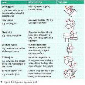
| Describe a joint's role with bones | a point in which bones connect, allow movement, provides support for bones, three types |
| Describe the structural and functional properties of a fibrous joint | held together by dense connective tissue, no movement permitted, visible lines, fuse later on -skull |
| Describe the structural and functional properties of a cartilaginous joint | connected by hyaline cartilage, provide relative movement, makes up the growth plate -vertabrae |
| Describe the structural and functional properties of a synovial joint | freely moving joint, contains synovial fluid to reduce shock, articular cartilage on epiphysis to protect joint during friction, susceptible to arthritis -knee |
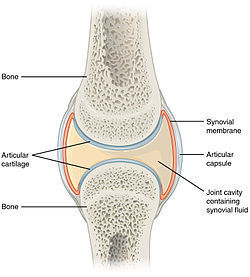
| (Diagram of a Synovial Joint-Knee) | |
| Describe the components of a synovial joint: Describe articular cartilage | smooth, white layer to protect against friction and absorb shock, wear + tear can be painful |
| Describe the components of a synovial joint: Describe synovial membrane and synovial fluid | the membrane creates the synovial fluid, a viscous liquid that provides nutrients to cartilage and reduces friction |
| Describe the components of a synovial joint: Describe bursae | fluid filled sacs, occur where two structures may rub together and areas of high stress (knees, elbows), lined with synovial membrane, reduces friction |
| Describe the components of a synovial joint: Describe the meniscus | discs of fibrocartilage, found between articulating bones, allow ill-fitting bones to better fit together, provides support and cushioning |
| Describe the components of a synovial joint: Describe the articular capsule | encapsulates the pieces of a synovial joint, allows joint movements |
| Name the 5 types of synovial joints | |
| Name and define the three characteristics of muscle tissue | Contractibility: ability to shorten/lengthen Extensibility: ability of muscle to stretch past it's normal length Elasticity: ability to return to oiginal resting length after stretch is removed |
| Name and describe the two types of changing muscle charactersitics (hint: they're opposites) | atrophy: decreasing size of muscle (old age, sickness) hypertrophy: increasing size of muscle (working out, puberty) |
| What is healthy muscle tissue controlle and fed by? | controlled by nerve stimuli fed by capillaries |
| Name the three types of muscle | Smooth muscle: involuntary, not striated, lines blood vessels walls, stomach, eyes, veins, trachea Cardiac muscle: involuntary control, striated, only in heart Skeletal muscle: voluntary control, striated, tendons connect it to bone, main function to move skeleton, quads, biceps |
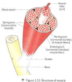
| (Muscle Fibre Diagram) | |
| Name the outer layer of skeletal muscle | Epimysium; covers all |
| What surrounds the bundles of muscle fibres? | Perimysium |
| What surrounds the individual muscle fibres? | Endomysium |
| Define Myofibril | bundles of actin + myosin inside muscle fibres |
| Define the characteristics and functions of a sarcomere | basic unit of a myofibril, forms long chains within the myofibril |
| Define Z-lines | indicate myofibril boundaries |
| Describe 'actin' strands | 'pearl' strands of proteins, helped by tropomyosin (bond with myosin) |
| Describe 'myosin' filaments | 'gold club' filaments, contains a head, a hinge and a rod, the head contains the ATP break down for energy |
| Define 'origin' of muscle | origin: the attachment of a muscle tendon to a stationary bone |
| Define 'insertion' of muscle | insertion: the attachgment of a muscle tendon to a moveable bone |
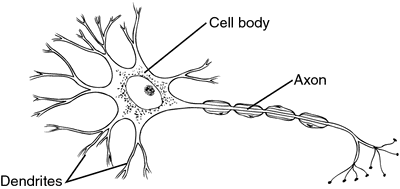
| Identify the components of a motor unit: Describe a dendrite | link to neuron through other neurons, allow info to flow between nerves |
| Identify the components of a motor unit: Describe a cell body | contained within the spinal cord or in clusters just outside it |
| Identify the components of a motor unit: Describe a nucleus | centre of cell body, contains all info for the cell |
| Identify the components of a motor unit: Describe an axon | main component on signal transmission |
| Identify the components of a motor unit: Describe a motor end plate | neuromuscular junction where the neuron joins the muscle fibre |
| Identify the components of a motor unit: Describe a synapse | the gap between the neuron and muscle fiber, neurotransmission travels, signal to stimulates muscle is sent |
| Neurotransmitter: Define Acetylcholine (Ach) | released in the synapse binding the muscle fiber and sending an action potential along the muscle fibers, begins action |
| Neurotransmitter: Define Cholinesterase/Acetylcholine esterase (Achase) | removes acetylcholine from the synapse, stops action |
| Describe the 3 types of muscle fibres | Type I: slow twitch, low force, high endurance, postural muscles, jogging Type IIa: fast twitch, medium force, medium endurance, swimming Type IIb: short bursts of power, weightlifting, jumping |
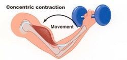
| Define the components of an isotonic muscular contraction | muscle shortens, movement occurs |
| Define the isotonic contraction: concentric contraction | muscle shortens as it contracts, ends of muscle draw together, flexion |
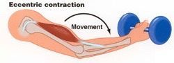
| Define the isotonic contraction: eccentric contraction | muscle lengthens as it contracts, pushing up, extension |
| Define isometric contraction | static exercise, no movement, shaking |
| Define isokinetic contraction | specialized movement, used for rehab, constant speed, pressure applied throughout movement, requires specialized equipment |
| Define reciprocal inhibition and it's two opposing muscle types | when one muscle contracts, the muscle with the opposing action relaxes agonist muscle: prime mover (bicep curl: bicep) antagonist muscle: relaxes in opposition (bicep curl: triceps) |
| Describe the causes of, the characteristics of treatments for DOMS (Delayed Onset Muscle Soreness) | caused by eccentric muscle contractions, the result of microscopic muscle damage, side effect of the repair process, mild muscle strain injury + inflammation, greatest pain 24-72-hour post exercise, treated with light activity, ice packs, manual massage, accupressure |
Want to create your own Flashcards for free with GoConqr? Learn more.