11963430

| Question | Answer |
| Define magnification | A measurement of the number of times larger an image is compared to its actual size Magnification = Image size / Object size |
| How many mm are there in one nanometer (nm)? | 0.000001 mm |
| How many mm are there in one micrometer (µm)? | 0.001 mm |
| Define resolution | How detailed the image is. Specifically, how well a microscope distinguishes between two points that are close together |
| What are the 3 types of microscope? | Light microscope Laser scanning confocal microscope Electron microscope |
| What are the two types of electron microscope? | Transmission electron microscope (TEM) Scanning electron microscope (SEM) |
| What are the properties of light microscopes? | Uses light Lower resolution than electron microscope Usually used to look at whole cells/tissues Maximum magnification = x1500 Maximum resolution = 0.2 µm |
| What are the properties of laser scanning confocal microscopes? | Uses laser beams to scan a specimen that is tagged with fluorescent dyes Can be used to look at objects at different depths in thick specimens Multiple images can be combined by the computer to generate 3D images |
| How does a laser confocal microscope produce an image? | 1. A laser beam is focused through a lens which is aimed at a beam splitter 2. This splits the beam and some of the light is directed to the specimen 3. This light is then focused through a pinhole onto a detector 4. The pinhole means that any out-of-focus light is blocked, producing a much clearer image than a normal light microscope 5. The detector is hoked up to a computer, which generates an image |
| What are the properties of electron microscopes? | Uses electrons instead of light Have a higher resolution so give more detailed images |
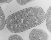
| What are the properties of transmission electron microscopes (TEMs)? | Uses electromagnets to focus a beam of electrons, which is then transmitted from the specimen to produce 2D images Denser parts of the specimen absorb more electrons, making it look more darker on the image Provides high resolution images to look at very small organelles Used to look at the internal structures of organelles in detail Specimens viewed must be thinly sliced |
|
What type of electron microscope generated this image?
Image:
Image (binary/octet-stream)
|
Transmission electron microscope (TEM) |
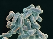
| What are the properties of scanning electron microscopes (SEMs)? | Scan a beam of electrons across the specimen This knocks off electrons from the specimen, which are gathered in a cathode ray tube to form an image The images produced show the surface of the specimen and can be 3D Gives lower resolution images than TEMs |
|
What type of electron microscope generated this image?
Image:
Image (binary/octet-stream)
|
Scanning electron microscope (SEM) |
| What is the maximum resolution and magnification of a light microscope? | LIGHT MICROSCOPE Resolution = 0.2 µm Magnification = x1500 |
| What is the maximum resolution and magnification of a TEM? | TEM Resolution = 0.0002 µm Magnification = Can be > x1,000,000 |
| What is the maximum resolution and magnification of a SEM? | SEM Resolution = 0.003 µm Magnification = Usually < x500,000 |
| Name 2 stains commonly used in light microscopy | Methylene blue Eosin |
| How does staining work? | The stain is taken up by some parts of the object more than others, meaning that some parts become more heavily stained than others. The contrast between heavily stained and lightly stained parts means that different parts of cells can be seen. |
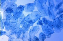
| What type of cell does methylene blue stain? |
DNA
(Above = cheek cell stained)
Image:
Image (binary/octet-stream)
|
| What types of cells do Giemsa stain differentiate? |
Commonly used to differentiate between different types of blood cells
(Above = blood smear stained)
Image:
Image (binary/octet-stream)
|
| Outline the process of using a light microscope (dry mount) | 1. Use tweezers to pick up your specimen and put it into the middle of a clean slide 2. Pop a cover slip on top |
| Outline the process of using a light microscope (wet mount) | 1. Pipette a small drop of the liquid onto the slide 2. Use tweezers to place the specimen on top of the drop 3. To put a cover slip on, stand the slip upright on the slide, next to the droplet 4. Carefully tilt and lower the slip so it covers the specimen (try not to get air bubbles underneath) 5. Once the cover slip is in position, you can add a stain. Put a drop of stain next to one edge of the cover slip and put a bit of paper towel next to the opposite edge 6. The stain will get drawn under the slip, across the specimen |
| How is an eyepiece graticule and stage micrometer used? | An eyepiece graticule (a transparent ruler with numbers, but no units) is fitted onto the eyepiece. The stage micrometer is placed on the stage - it is a microscopic slide with an accurate scale (it has units) and is used to work out the value of the divisions on the eyepiece graticule at a particular magnification. |
Want to create your own Flashcards for free with GoConqr? Learn more.