
Description
Page 1
Schematic model of the molecular mechanisms that regulate ionotropic glutamate receptor distribution during hippocampal mGluR-LTD induction and expression. Activation of group I mGluRs leads to dephosphorylation and activation of STEP, which transiently dephosphorylates tyrosine (Y) residues in GluA2-containing AMPARs at the PSD. This may trigger lateral diffusion of AMPARs to extrasynaptic endocytic zones from which internalization occurs. It is likely that upon internalization, AMPARs are rephosphorylated by PTKs and retained at intracellular compartments until recycled or degraded. In parallel to STEP activation, group I mGluR activation can stimulate ERK1/2 and p38 MAPK signaling pathways via G-protein release and activation of the small GTPase Rap. ERK1/2 activation could stimulate calpain proteolysis of NMDAR and AMPAR subunit C-termini, which facilitates loss of synaptic receptors. Protease involvement is also exemplified by TACE, which cleaves NPR and allows it to cluster with AMPARs and other NPs in endocytic vesicles. Activation of the p38 MAPK signaling pathway stimulates the formation of the GDI-Rab5 complex, which also mediates AMPAR endocytosis. mGluR-LTD involves regulation of gene transcription and protein translation via the activation of signaling pathways such as p38 MAPK and ERK. Protein synthesis up-regulated includes STEP, FMRP, Arc/Arg3.1, and MAP1B, which are all involved in regulation of AMPAR internalization. After synthesis, FMRP is rapidly degraded such that it can no longer inhibit translation of specific mRNAs such as Arc/Arg3.1 and can no longer down-regulate AMPAR endocytosis. Arc/Arg3.1 forms a complex with endophilin2/3 (Endo) and dynamin (Dyn), which facilitates AMPAR internalization. MAP1B also promotes receptor endocytosis by sequestering the scaffolding protein GRIP away from the PSD such that AMPAR synaptic stabilization is reduced. The dotted line corresponds to a postulated pathway involved in mGluR-LTD, solid arrows relate to confirmed molecular mechanisms that promote AMPAR internalization, and the inhibitory arrows refer to pathways that are down-regulating protein synthesis or receptor endocytosis. Figure is based on data described in Zhang et al. (2008)and Gladding et al. (2009).
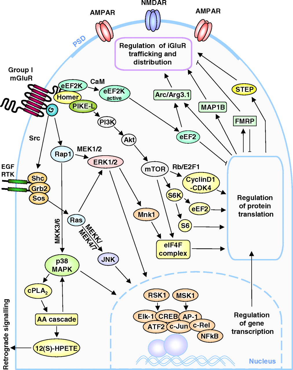
Schematic model of molecular mechanisms underlying regulation of gene transcription and protein translation during mGluR-LTD in postsynaptic neurons of hippocampal CA3-CA1 synapses. Group I mGluR stimulation can activate the epidermal growth factor receptor tyrosine kinase (RTK) via G-protein release, which activates the nonreceptor tyrosine kinase Src. Recruitment of the Shc-Grb2-Sos complex stimulates the small GTPase Ras, which leads to activation of ERK1/2 and JNK MAPK signaling cascades. ERK1/2 and p38 MAPK can also be stimulated by G-protein release via the small GTPase Rap1 through the mitogen-activated protein kinase kinases MKK3/6 and MEK1/2, respectively. Stimulation of ERK1/2, JNK, and p38 MAPK signaling cascades leads to activation of the transcription factors Elk-1, CREB, activator protein-1 (AP-1), ATF2, c-Jun, c-Rel, and NF-κB via the kinases RSK1 and mitogen and stress-activated protein kinase-1. p38 MAPK is also linked to activation of the AA cascade of which the 12(S)-HPETE metabolite may serve as a retrograde signaling molecule. In addition, ERK1/2 signaling can mediate formation of the translation initiation eIF4F complex necessary for initiating protein synthesis. mGluR-LTD involves activation of the PI3K-Akt-mTOR signaling pathway via interactions with Homer and PI3K enhancer-L (PIKE-L). This can facilitate regulation of protein synthesis by activating S6K, eEF2, the eIF4F complex, and the cyclin1-CD4 complex. Furthermore, mGluR activation leads to dissociation and activation of eEF2K, which phosphorylates eEF2. Activated eEF2 prevents global protein translation but facilitates up-regulation of Arc/Arg3.1 synthesis, which is involved in AMPAR endocytosis. Other proteins synthesized during mGluR-LTD include STEP, FMRP, and MAP1B, which may also regulate AMPAR synaptic trafficking.
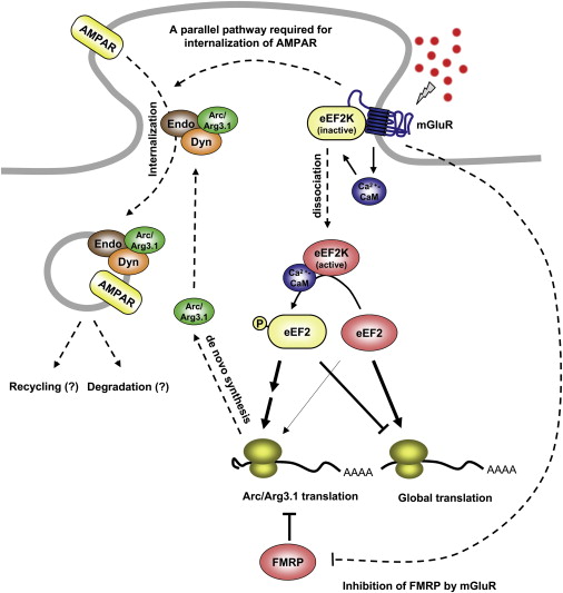
Figure 8. eEF2K, FMRP, and Rapid De Novo Translation of Arc/Arg3.1 Protein in mGluR-LTDGroup I mGluRs activate eEF2K via calcium-calmodulin (CaM). eEF2K phosphorylates eEF2, which inhibits elongation generally but increases Arc/Arg3.1 translation. Arc/Arg3.1 forms a complex with endophilin2/3 (Endo) and dynamin (Dyn) and induces the internalization of AMPAR (Chowdhury et al., 2006). FMRP inhibits the translation of Arc/Arg3.1 at the basal state. Arc/Arg3.1 induction alone is not sufficient for mGluR-LTD, indicating that mGluR activates another pathway that is required to internalize AMPAR (Cho et al., 2008). In Fmr1 KO mice, the synthesis of Arc/Arg3.1 protein is constitutively derepressed, and de novo synthesis of Arc/Arg3.1 is not required for mGluR-LTD.
Signalling
gene expression
FMRP signalling
0 comments
Want to create your own Notes for free with GoConqr? Learn more.