Description
|
|
Created by Cher Bachar
over 11 years ago
|
|
Page 1
Schematic connections between the pre-frontal cortex and limbic structures within the limbic-cortico-striato-pallido-thalamic circuits related to the medial and orbital prefrontal cortex networks implicated in depression. A decrease in the inhibitory control of the limbic structures by the PFC is associated with cognitive, behavioural and other signs of depression as well as abnormalities in neuroendocrine function, pain modulation and neurotransmitter activity (affecting the raphe, serotonergic nuclei and NA-ergic nucleus coeruleus), through its connections with the hypothalamus and the midbrain, in particular the periaqueductal area.
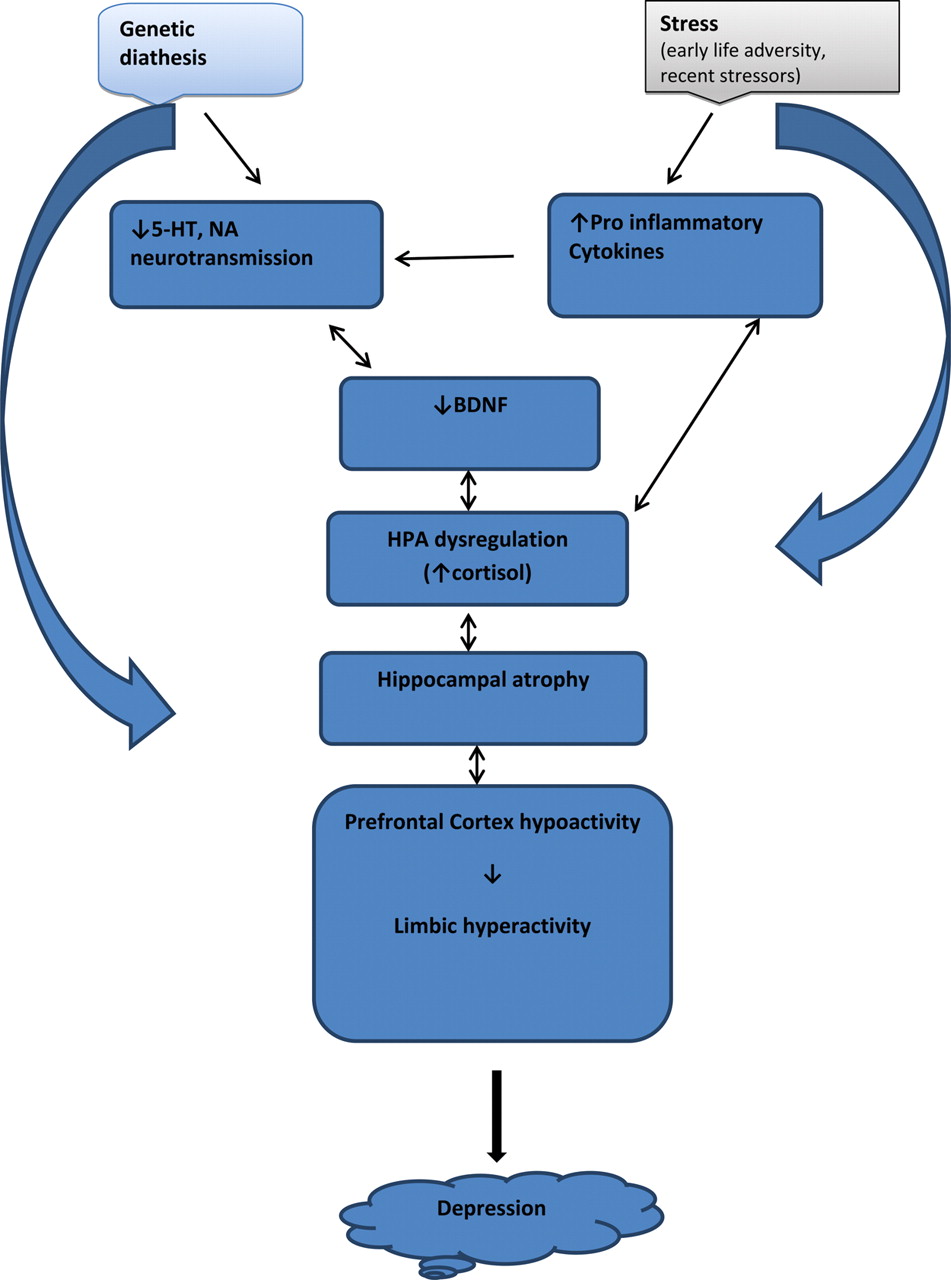
The structural changes in the brain in particular the hippocampus and PFC are believed to be due to abnormalities in neuroplasticity rather than neurodegeneration. Nevertheless, it remains to be confirmed whether these changes are indeed always reversible, particularly in the PFC and also whether or not they predate the onset of depression. The dysregulation of the HPA axis is responsible to a great extent for these abnormalities. Raised levels of circulating cortisol activate brain receptors stimulating gene transcription and protein synthesis. Although this may have a beneficial effect in the short term, enabling the brain to cope with smaller amounts of stress, persistent hypercortisolaemia in chronic stress can affect voltage-gated ion channels allowing increased calcium entry into the activated neurons and causing neuronal damage. Glucocorticoid-induced damage in the hippocampus may occur directly, via activation of the glutamate systems or via BDNF reduction. CRH also has direct toxic effects on hippocampal neurons. Stress is associated with reduced BDNF concentrations which further impair neuronal survival. The decrease in BDNF concentrations may be due to the reduction in hippocampal neuronal tissue, as well as a direct effect of hypercortisolaemia; decreased activity in monoaminergic neurotransmission or other noxious factors (?glutamate effects) may also be responsible. The resulting impaired hippocampal function fails to adequately regulate (inhibit) the HPA axis, therefore sustaining ‘toxic’ hypercortisolaemia. The rise in cytokines levels may also contribute to the sustained HPA activation and abnormal IRSs may have a secondary or even a primary role in the dysregulation of the HPA axis.
animal models
functional and structural changes in depression
neuroanatomical connectivity
chemical imbalance
Want to create your own Notes for free with GoConqr? Learn more.