903989
Description

Mind Map by toronto416, updated more than 1 year ago
|
|
Created by toronto416
over 10 years ago
|
|
MCAT Review:
Human Skeletal
System
- Other
- 206 bones
- Types of Bones
- Long
- Short
- Flat
- Irregular
- Long
- Types of Bones
- Cartilage
- Tendons
- Ligaments
- 206 bones
- Axial Skeleton
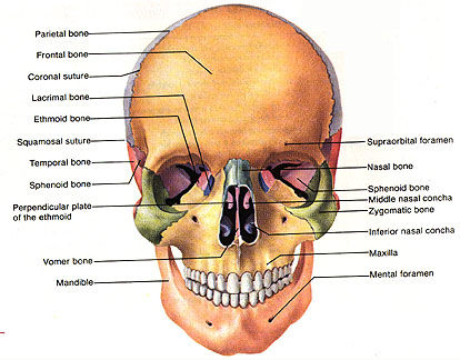
- Skull
- Cranial
- Etmoid
- Frontal
- Occipital
- Parietal
- Sphenoid
- Temporal
- Etmoid
- Facial
- Lacrimal
- Mandible
- Maxilla
- Nasal
- Palatine
- Turbinate
- Vomer
- Zygomatic
- Lacrimal
- Cranial
- Sternum
Annotations:
- The sternum is the flat bone located in the middle of the chest. The sternum, together with the ribs, forms the ribcage that helps to protect the heart and the lungs, as well as major blood vessels. Ribs are connected to the sternum by cartilage. The sternum is composed of the following three parts: the manubrim, the gladiolus and the xiphoid process. In adults, the three parts of the sternum are often bone that is fused together.
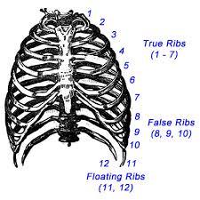
- Ribs
- True Ribs
- False Ribs
- Floating Ribs
- True Ribs
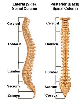
- Vertebral Column
- Cervical Vertebrae
- Thoracic Vertebrae
- Lumbar Vertebrae
- Sacrum
- Coccyx
- Cervical Vertebrae
- Skull
- Appendicular
Skeleton
- Upper
Extremeties
- Arm
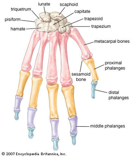
- Hand
- Forarm
- Arm
- Low Extremeties
- Thigh
Annotations:
- The only bone in the thigh is the femur. It extends from the hip to the knee and is the largest and strongest bone in the body. The femur connects to the kneecap to form the knee (together with the tibia).
- Leg
Annotations:
- The leg is made up of two bones, the tibia and the fibula. These bones extend from the knee to the foot. The fibula is located on the outside of the leg and is the smaller of the two bones in the leg. The fibula's primary purpose is muscle attachment. The tibia is on the inside of the leg and is also called the "shin" bone. It connects to the patella to form the knee, and it connects to the foot, together with the talus (anklebone) to assist with flexing and extending the foot. The tibia bears most of the weight in the leg portion of the lower extremities.
- Patella
Annotations:
- The patella is a sesamoid bone that sits between the femur (thigh) and the tibia (leg). It helps to protect the knee joint, and also helps to strengthen the tendons in the knee.
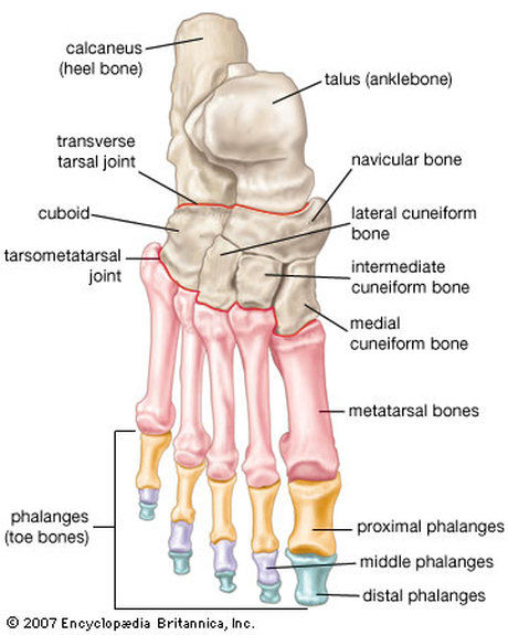
- Foot
Annotations:
- The foot (also called the "pes") is made up of 26 bones that are divided into three parts: the ankle (tarsus), the instep (metatarsus), and the toes (phalanges). The bones of the ankle (tarsus) includes seven tarsal bones, similar to the carpal bones in the wrist. The diagram below shows the placement and names of all seven tarsal bones. The largest of all of the tarsal bones is the calcaneous bone, which is also called the "heel." There are five metatarsal bones that make up the instep of the foot. They are numbered I-V beginning on the inside of the foot. The metatarsal bones are in the same location in the foot as the metacarpal bones in the hands. The phalanges in the foot are arranged in the same fashion as the phalanges in the hand. The diagram below shows the placement of all of the bones in the foot.
- Thigh
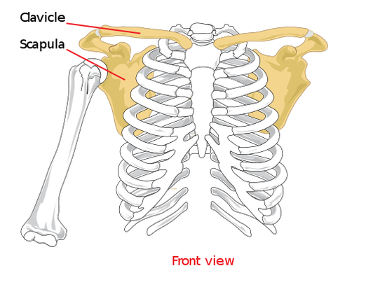
- Shoulder Girdle
- Clavicles
Annotations:
- The clavicle is the bone in the shoulder girdle that is often referred to as the collarbone. It is a curved bone that connects the upper extremity (arm) to the body (the trunk), and provides several attachment sites for muscles and ligaments. The clavicle is unique in that a portion of this bone is not covered by muscle, but only covered by skin. Because of this difference, it is oven visible on many people. The clavicle's primary purpose is to maintain distance between the upper extremity and trunk to allow the shoulder to move freely.
- Scapulea
Annotations:
- The scapula is the bone in the shoulder that is often referred to as the shoulder blade (because of its blade-like appearance). The primary purpose of the scapula is to connect the humerus (upper arm bone) to the clavicle (collar bone). The scapula forms the back portion of the shoulder girdle and has several ridges on the front and back that allow muscles, tendons and ligaments to attach. Below is a diagram that shows the clavicles and the scapulae.
- Clavicles
- Pelvic
Girdle
Annotations:
- The pelvic girdle, which is also referred to as the hip girdle, consists of two hip bones (left and right). The hip bones are also called the coxal bones. At birth the coxal bones are divided into three parts: the ilium, the ischium, and the pubis. As one ages, the three bones fuse together. The two hip bones meet in the back on either side of the sacrum. In the front the two bones are connected by a muscle. The pelvic girdle serves several purposes in the body. First, it, together with the vertebral column, helps to support the body's weight and maintain the body upright. The pelvic girdle also protects the important organs of the urinary system and reproductive systems, and in a female protects a devloping fetus during pregnancy. One thing that is unique about the pelvic girdle is that it differs in males and females. In males, the pelvic girdle is larger and the bones are closer together. In a female the pelvic girdle is smaller, but the bones are further apart to allow for a fetus to pass through during childbirth. The diagram below shows the pelvic girdle.
- Upper
Extremeties
Media attachments
Want to create your own Mind Maps for free with GoConqr? Learn more.