9138730
Description
Flashcards by Victoria Wright, updated more than 1 year ago
|
|
Created by Victoria Wright
over 7 years ago
|
|
| Question | Answer |
| What is the first phase of the left ventricle? | Isovolumetric contraction—period between mitral valve closing and aortic valve opening; period of highest O2 consumption |
| Describe the Isovolumetric contraction phase of the left ventricle? | 1) Isovolumetric contraction—period between mitral valve closing and aortic valve opening; period of highest O2 consumption |
| Which phase of the left ventricle has the highest O2 consumption? | Isovolumetric contraction—period between mitral valve closing and aortic valve opening; period of highest O2 consumption |
| What is the second phase of the left ventricle? | Systolic ejection—period between aortic valve opening and closing |
| What is the third phase of the left ventricle? | Isovolumetric relaxation—period between aortic valve closing and mitral valve opening |
| What is the fourth phase of the left ventricle? | Rapid filling—period just after mitral valve opening |
| What is the fifth stage of the left ventricle? | Reduced filling—period just before mitral valve closing |
| Describe the Systolic ejection phase of the left ventricle? | 2) Systolic ejection—period between aortic valve opening and closing |
| Describe the Isovolumetric relaxation phase of the left ventricle? | 3) Isovolumetric relaxation—period between aortic valve closing and mitral valve opening |
| Describe the rapid filling phase of the left ventricle? | 4) Rapid filling—period just after mitral valve opening |
| Describe the reduced filling phase of the left ventricle? | Reduced filling—period just before mitral valve closing |
| Heart sounds: Where is the S1 loudest? | S1—mitral and tricuspid valve closure. Loudest at mitral area. |
| Heart sounds. What sound is described below? Mitral and tricuspid valve closure | S1 |
| Heart sounds. What sound is described below? Aortic and pulmonary valve closure. | S2 |
| Heart sounds. What sound is described below? Loudest at left upper sternal border. | S2 |
| Heart sounds. What sound is described below? Loudest at mitral area | S1 |
| Heart sounds. What sound is described below? In early diastole during rapid ventricular filling phase | S3 |
| Heart sounds. What sound is described below? Associated with increased filling pressures | S3 |
| Heart sounds. What sound is described below? Associated with mitral regurgitaion, HF | S3 |
| Heart sounds. What sound is described below? More common in dilated ventricles | S3 (but can be normal in children and young adults) |
| Heart sounds. What sound is described below? In late diastole (“atrial kick”) | S4 |
| Heart sounds. What sound is described below? Best heard at apex with patient in left lateral decubitus position. | S4 |
| Heart sounds. What sound is described below? High atrial pressure. | S4 |
| Heart sounds. What sound is described below? Associated with ventricular noncompliance | S4 |
| Heart sounds. What sound is described below? Associated with hypertrophy | S4 |
| Heart sounds. What sound is described below? Left atrium must push against stiff LV wall. | S4 |
| Heart sounds. What sound is described below? Consider abnormal, regardless of patient age. | S4 |
| Heart sounds: Where is S2 loudest? | Loudest at left upper sternal border. |
| Heart sounds: Where is S4 best heard? | Best heard at apex with patient in left lateral decubitus position. |
| Describe S1 heart sounds. | S1—mitral and tricuspid valve closure. Loudest at mitral area. |
| Describe S2 heart sound. | S2—aortic and pulmonary valve closure. Loudest at left upper sternal border. |
| Describe S3 heart sound. | S3 |
| Describe S4 heart sound. | S4 - in late diastole (“atrial kick”). Best heard at apex with patient in left lateral decubitus position. High atrial pressure. Associated with ventricular noncompliance (eg, hypertrophy). Left atrium must push against stiff LV wall. Consider abnormal, regardless of patient age. |
|
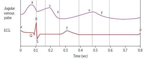
Jugular venous pulse
Describe (a)
Image:
Capture (image/jpeg)
|
(a) wave - atrial contraction. Absent in atrial fibrillation (AF). |
|
Jugular venous pulse
Describe (c)
Image:
Capture (image/jpeg)
|
(c) wave—RV contraction (closed tricuspid valve bulging into atrium). |
|
Jugular venous pulse
Describe (x)
Image:
Capture (image/jpeg)
|
(x) descent—downward displacement of closed tricuspid valve during rapid ventricular ejection phase. Reduced or absent in tricuspid regurgitation and right HF because pressure gradients are reduced. |
|
Jugular venous pulse
Describe (v)
Image:
Capture (image/jpeg)
|
(v) wave—increased right atrial pressure due to filling (“villing”) against closed tricuspid valve. |
|
Jugular venous pulse
Describe (y)
Image:
Capture (image/jpeg)
|
(y) descent—RA emptying into RV. Prominent in constrictive pericarditis, absent in cardiac tamponade. |
| JVP. Which is absent in atrial fibrillation? |
(a) wave
Image:
Capture (image/jpeg)
|
| JVP. Which is reduced or absent in tricuspid regurgitation and right HF? Why? |
(x) decent - Reduced or absent in tricuspid regurgitation and right HF because pressure gradients are reduced.
Image:
Capture (image/jpeg)
|
| JVP. Which is absent in cardiac tamponade? |
(y) decent
Image:
Capture (image/jpeg)
|
| JVP. Which is prominent in constructive pericarditis? |
(y) decent
Image:
Capture (image/jpeg)
|
Want to create your own Flashcards for free with GoConqr? Learn more.