1922556
Description
Flashcards by Alice Rabone, updated more than 1 year ago
|
|
Created by Alice Rabone
over 9 years ago
|
|
| Question | Answer |
| Define 'eukaryote' | Complex cells which have a true nucleus and other membrane bound organelles. |
| Define 'prokaryotic' | Smaller, simpler cells which don't have a true nucleus or membrane bound organelles. |
| List the organelles found in an animal cell. | Plasma membrane, RER,SER, nucleus, nucleolus, lysosomes, ribosomes, nuclear envelope, Golgi apparatus, cytoplasm and mitochondria. |
| Which extra organelles do plant cells have that animal cells don't have? | A cell wall (cellulose) with plasmodesmata, a vacuole, chloroplasts. |
| What is the structure and function of the plasma membrane? | The membrane found on the surface of animal cells and just under the cell wall in plant cells and prokaryotic cells, made mainly of proteins and lipids. It regulates the movement of substances in and out of then cell and has receptor molecules on it to allow response to chemicals such as hormones. |
| What is the structure and function of the cell wall? | A rigid structure that surrounds plant cells, made of cellulose- also found in some bacteria cells but made from peptidoglycan, not cellulose. |
| What is the structure and function of the nucleus? | A large organelle surrounded by a double membrane called the nuclear envelope, containing many pores. The nucleus contains chromatin, which is made from proteins and DNA, and the nucleolus. The DNA controls the cell's activities. The nuclear pores allow substances (e.g RNA) to move between the nucleus and cytoplasm. The nucleolus makes ribosomes. |
| What is the structure and function of lysosomes? | A round organelle surrounded by a membrane, with no clear internal structure. Contains digestive enzymes, kept separate from the cytoplasm by the membrane, which can be used to digest invading cells or break down worn out components in a cell. |
| What is the structure and function of ribosomes? | Very small organelles that either float free in the cytoplasm or is attached to the RER. Proteins are made at the ribosomes. |
| What is the structure and function of the rough endoplasmic reticulum? | A system of membranes enclosing a fluid-filled space. The surface is covered in ribosomes. Its function is to fold and process proteins made at the ribosomes. |
| What is the structure and function of the smooth endoplasmic reticulum? | A system of membranes enclosing a fluid filled space (like RER but without ribosomes). Its function is to synthesise and process lipids. |
| What is the structure and function of vesicles? | A small fluid filled sac in the cytoplasm, surrounded by a membrane, which transports substances in and out of the cell (via the plasma membrane) and between organelles. Some are formed at the Golgi Apparatus or the ER, while others are formed at the cell surface. |
| What is the structure and function of the Golgi apparatus? | A group of fluid filled, flattened sacs, often surrounded by vesicles. It processes and packages new lipids and proteins, and makes lysosomes. |
| What is the structure and function of mitochondria? | Usually oval shaped and have a double membrane- the inner one is folded to form structures called cristae. Inside is the matrix, which contains enzymes involved in respiration to produce ATP. They are found in large numbers in cells very active cells which require lots of energy. |
| What is the structure and function of chloroplasts? | A small flattened structure found in plant cells, surrounded by a double membrane and also has membranes inside called thylakoid membranes. These membranes are stacked up to form grana. They are the site of photosynthesis- some of the process takes place in the grana and some in the stroma (a thick fluid in chloroplasts). |
| What is the structure and function of the centrioles? | Small hollow cylinders containing a ring of microtubules (tiny protein cylinders). The centrioles are involved with the separation of chromosomes during cell division. |
| What is the structure and function of the cilia? | Small hair-like structures found on the surface of some animal cells. The microtubules inside allow the cilia to move, to move substances along the cell surface. |
| What is the structure and function of flagellum? | Like cilia, they have 2 microtubules in the centre and 9 pairs around the edge, and stick out from the cell surface. The microtubules contract to make the flagellum move, like an outbound motor to propel the cell forward. |
| What is the equation for magnification? | Magnification= image size/actual size |
| Define 'magnification'. | The degree to which an image is larger than the actual specimen size. |
| Define 'resolution'. | The smallest distance between two points on a specimen that can still be distinguished as two separate entities. |
| What are the main features of a light microscope? | They use light Have a maximum resolution of 200nm They have a maximum useful magnification of only x1500 |
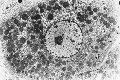
| What are the main features of a transmission electron microscope? | Use electrons instead of light to form an image. They have a resolution of around 0.1 nm, and a magnification of x1,000,000. They use electromagnets to focus a beam of electrons, which make them transmitted through the specimen- denser parts of the specimen absorb more electrons so have a darker appearance on the image. |
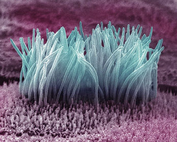
| What are the main features of a scanning electron microscope? | Use electrons instead of light to form an image. They have a resolution of around 5nm, and a magnification of just under x1,000,000. They scan a beam of electrons across the specimen. This knocks off electrons from the surface of the specimen, which are gathered in a cathode ray to give a 3D image. |
| What type of microscope produced this image? | TEM image of human liver |
| What type of microscope would produce this image? | An SEM image of cilia cells in a human lung. |
| What is meant by 'staining'? | Adding a dye (light microscopes) or dipping objects into a solution of heavy metals (like lead), in order to give the image produced contrast. |
| What is the purpose of cell surface membranes? | They control which substances enter and leave the cell, as they are partially permeable. They allow recognition by other cells e.g. the cells of the immune system. They allow cell communication. They define the cell's perimeter. |
| What is the purpose of membranes within cells? | To divide the cell into compartments, to make processes more efficient. Folded membranes inside cells increase surface area, which increases efficiency of some reactions e.g. respiration. They can form vesicles for transport. To control which substances enter and leave the organelle- e.g the nuclear membrane, which is partially permeable. |
| Describe the fluid mosaic model. | Phospholipid bilayer 'Fluid' because bilayer is always moving. Cholesterol gives stability. Scattered protein molecules. Some proteins have polysaccharide chains attached- called glycoproteins. Some lipids also have polysaccharide chains attached- called glycolipids. |
| What role do the phospholipids have in a cell membrane? | Hydrophilic head Hydrophobic tail Arranged into a bilayer with heads facing outwards. The centre of the bilayer is hydrophobic so doesn't allow water soluble substances (like ions) through it, acting as a barrier to these dissolved substances. |
| What role does cholesterol play in cell membranes? | A type of lipid. Fits between the phospholipids. Binds to the hydrophobic tails, causing them to pack more closely together, making the membranes less fluid and more rigid. |
| What is the role of proteins in cell membranes? | Control what enters and leaves the cell. Some form channel proteins and others form carrier proteins. Proteins also act as receptors for molecules in cell signalling- when a molecule binds to the protein a chemical reaction is triggered inside the cell. |
| What roles do glycoproteins and glycolipids have in cell membranes? | Stabilise the membrane by forming hydrogen bonds with surrounding water molecules. Sites where drugs, hormones and antibodies bind. They act as receptors for cell signalling. They are antigens involved in the immune response. |
| How do cells communicate with each other? | One releases a messenger molecule (e.g. a hormone), which travels in the blood to another cell, where it is detected when it binds to a receptor on the plasma membrane. |
| Why do receptor molecules have specific shapes? | They have complementary shapes to their corresponding messenger molecules. A cell which responds to a particular messenger molecule is referred to as a target cell. |
| How do many drugs work? | By binding to receptors on cell surface membranes. This either triggers a response or blocks the receptor to prevent it from working. |
| How are membranes affected by temperature? | Increase in temperature = increase in fluidity. Increase in kinetic energy of molecules. Increase in permeability of membrane. Decrease in temperature = decrease in fluidity. Decrease in kinetic energy of molecules. Decrease in permeability of membrane. Unsaturated fatty acids increase fluidity. Organisms in cold environments need more unsaturated fatty acids to increase fluidity. |
| What is diffusion? | The passive, net movement of particles from an area of high to low concentration, down a concentration gradient. Diffusion continues until an equilibrium is reached. |
| What factors affect the rate of diffusion? | The concentration gradient- the greater the difference, the faster the rate. Diffusion distance Surface area |
| What is osmosis? | The diffusion of water molecules from a high to low water potential. Water potential is the likelihood of water molecules to diffuse into/out of a solution. Usually a negative number, except pure water, which has a WP of 0. |
| What happens to an animal cell/plant cell put into a hypotonic solution (higher water potential than cell)? | Animal cell: net movement of water into the cell, causing it to burst because the plasma membrane can't withstand the pressure. Plant cell: net movement of water into the cell, causing the vacuole to swell, so the cytoplasm pushes against the cell wall. The cell has a high pressure potential and is turgid. |
| What happens to an animal cell/plant cell put into a hypertonic solution (lower water potential than cell)? | Animal cell: net movement of water out of the cell, causing it to shrink (crenated). Plant cell: net movement of water out of the cell, causing the cell to become flaccid (limp). The plasma membrane and the cytoplasm pull away from the cell wall, known as plasmolysis. |
| What is facilitated diffusion? | The use of carrier/channel proteins. |
| What are channel proteins? | Form pores in the membrane for charged particles (ions) which cannot diffuse straight through the membrane. |
| What are carrier proteins? | Move larger molecules across membranes. Different carrier proteins facilitate the diffusion of different molecules. Firstly, a large molecule attaches to the carrier protein, which then changes shape to release the molecule on the other side of the membrane. |
| What is active transport? | The net movement of particles against a concentration gradient, across plasma membranes, which requires carrier proteins and energy (ATP). |
| What is endocytosis? Give an example. | Cells take in large substances by endocytosis, by surrounding it with a section of its plasma membrane, which pinches off to form a vesicle with the ingested substance inside. E.g. phagocytosis |
| What is exocytosis? Give an example. | How cells secrete substances such as digestive enzymes, hormones and lipids. Vesicles pinch off the Golgi apparatus and move to the plasma membrane, where they fuse and release their contents outside the cell. Some substances are released straight into the plasma membrane such as membrane proteins. |
| What is the cell cycle? | The process of cell growth and division, involving interphase and mitosis. |
| What is mitosis? | Cell replication to produce 2 genetically identical daughter cells. Needed for growth and repair. Has 4 stages. |
| What is interphase? | Comes before mitosis in the cycle- when the cell grows and replicates its DNA and organelles in preparation for division. |
| What is prophase? | The first stage of mitosis, where the chromosomes condense (get shorter and fatter so visible under microscope). The centrioles start to move to the poles to form the spindle, and the nuclear envelope begins to break down. |
| what happens in metaphase? | The chromosomes line up along the middle of the cell, joined to the spindle fibres by their centromeres. |
| What happens in anaphase? | The centromeres divide, separating the sister chromatids. The spindles contract, pulling chromatids to opposite ends of the cell by their centromeres. |
| What happens in telophase? | The chromatids reach opposite poles on the spindle, so they uncoil and become chromosomes again. A nuclear envelope forms around each group of chromosomes so there is now two nuclei. The cytoplasm divides, and cytokinesis follows to give 2 genetically identical daughter cells. |
| How can you observe mitosis? | By staining chromosomes and looking at the cells under a microscope it is possible to see the different stages of mitosis. |
| Describe the process of yeast budding. | A bud forms on the surface of the yeast cell. The cell undergoes interphase, where the DNA and organelles are replicated. The cell then begins to undergo mitosis, and when nuclear division is complete the budding cell contains a nucleus that has an identical copy of the parent cell's DNA. Finally the bud separates off from the parent cell, producing a clone. |
| What are stem cells? | Cells which have all of their genes 'switched on' and have the potential to differentiate into a specialised cell. They are found in bone marrow and early embryos. |
| What is the difference between embryonic stem cells and stem cells found in adults? | Embryonic stem cells can develop into any type of human cell. Adult stem cells can only develop into a limited range of cells e.g. blood cells. |
| What is the process of cells specialising called? | Differentiation- this is where certain, unwanted genes are 'turned off'. |
| Where are stem cells found in plants and what can they differentiate into? | In the cambium- these cells can become xylem or phloem cells. The vascular cambium forms a ring in the roots and shoots, and cells divide and grow out from the ring, differentiating as they move away from the cambium. |
| How is a neutrophil (animal cell) adapted for its function? | White blood cell to defend against disease:- flexible shape to engulf pathogens many lysosomes in their cytoplasm, containing digestive enzymes to digest engulfed particles. |
| How are erythrocytes (animal cell) adapted for their function? | Red blood cells to carry oxygen:- biconcave disc shape gives a large surface area and no nucleus means more space for haemoglobin, the protein which carries oxygen- binds to it to form oxyhemoglobin. |
| How are epithelial cells (animal cells) adapted for their function? | Cover the surfaces of organs:- cells joined by interlinking cell membranes and a membrane at their base. Some have cilia that beat to move particles away. Others have microvilli to increase the cell's surface area. |
| How are sperm cells (animal cells) adapted for their function? | Male gamete:- flagellum (tail) to propel towards egg lots of mitochondria for respiration, to provide energy to swim the acrosome contains digestive enzymes to allow the sperm cell to penetrate the egg during fertilisation |
| How are palisade mesophyll cells (plant cells) adapted for their function? | Found in leaves, where photosynthesis occurs:- contain lots of chloroplasts to absorb sunlight for photosynthesis thin cell walls- short diffusion distance for gases (CO2, O2) to get in/out |
| How are root hair cells (plant cells) adapted for their function? | Absorb water and mineral ions from soil:- large surface area thin permeable cell wall for easy entry of water and mineral ions cytoplasm contains extra mitochondria to release energy needed for active transport |
| How are guard cells (plant cells) adapted for their function? | Line the stomata:- in the light, guard cells take up water and become turgid. Their thin outer walls and thickened inner walls force them to bend outwards, opening the stomata. This allows the leaf to exchange gases for photosynthesis. |
| What is a tissue? Give examples. | A group of cells (and any extracellular material secreted by them) that are specialised to work together to carry out a particular function. Examples: -squamous epithelium tissue (lining) -xylem tissue (transports water+supports plant- contains xylem and parenchyma cells) -ciliated epithelium (moves substances e.g mucus in trachea) -phloem tissue (transports sugar, arranged into tubes and made up of sieve cells, companion cells and ordinary plant cells.) |
| What is an organ? | A group of tissues that work together to perform a particular function. Examples: -the lungs -leaves |
| What is an organ system? | A group of organs working together to perform a specific function. Examples: -respiratory system -circulatory system |
| Give examples of substances that an organism needs to exchange with the environment. | -Oxygen -Carbon dioxide -Nutrients -Water -Urea |
| Why do large organisms need a dedicated exchange system? | The larger the organism the smaller its surface area:volume ratio. Large organisms cannot rely on diffusion to provide all of its cells with substances such as glucose and oxygen because: -diffusion distances are large (some cells are deep inside the body) -diffusion is slow |
| Explain how the lungs are an exchange organ. | 1) As you breathe in, air enters the trachea 2) The trachea splits into 2 bronchi- one for each lung 3) Each bronchi then branches off into many bronchioles 4) The bronchioles enter the alveoli, where gases are exchanged 5) The ribcage, intercostal muscles and diaphragm all work together to move air in/out |
| How are the alveoli adapted for gas exchange? | 1) good blood supply 2) thin walls- short diffusion distance 3) moist surface 4) large surface area |
| What features does a good gas exchange surface have? | 1) large surface area 2) short diffusion distance- thin wall 3) steep concentration gradient maintained All increase the rate of diffusion. |
| What function do goblet cells play (in the respiratory system)? | They secrete mucus, which traps microorganisms and dust particles in inhaled air, stopping them reaching the alveoli. |
| What function do cilia cells play (in the respiratory system)? | They beat mucus up the trachea, away from the alveoli, to the throat where it can be swallowed. This helps prevent lung infections. |
| What function do elastic fibres play (in the respiratory system)? | Elastic fibres on the walls of the trachea, bronchi, bronchioles and alveoli help the process of breathing out. On breathing in the lungs inflated and elastic fibres are stretched. Then the fibres RECOIL to help with exhalation by pushing the air out. |
| What function does smooth muscle play (in the respiratory system)? | Allows the diameter of the trachea, bronchi and bronchioles to be controlled. During exercise the muscle relaxes, making the tubes wider so theres less resistance to air flow and more air can enter/leave the lungs. |
| What function do rings of cartilage play (in the respiratory system)? | Provides the walls of the trachea and bronchi with SUPPORT. It's strong but flexible- it stops the airways collapsing when you breathe in and pressure drops. |
| Describe the structure of the trachea | 1) Large C shaped rings of cartilage 2) Smooth muscle for expansion 3) Elastic fibres for recoil 4) Goblet cells to secrete mucus 5) Ciliated epithelium to beat mucus away from the lungs |
| Describe the process of inspiration. | 1) Intercostal muscles and diaphragm contract, moving the ribcage upwards and outwards 2) Thorax volume increases 3) Lung pressure decreases (to below atmospheric pressure) 4) Air flows into the lungs |
| Describe the process of exhalation. | 1) Intercostal muscles and diaphragm relax, moving the ribcage downwards and inwards 2) Thorax volume decreases 3) Lung pressure increases (to more than atmospheric pressure) 4) Air flows out of the lungs |
| What is tidal volume? | The volume of air in a normal breath- usually around 0.4 dm^3 |
| What is vital capacity? | The maximum volume of air that can be breathed in or out. |
| What is breathing rate? | How many breaths are taken, usually in a minute. |
| What is oxygen uptake? | The rate at which a person uses up oxygen (the number of dm^3 used per minute) |
| What does a spirometer machine give readings for? | Tidal volume Vital capacity Breathing rate Oxygen uptake |
Want to create your own Flashcards for free with GoConqr? Learn more.