441685
Description
Flashcards by gina_evans0312, updated more than 1 year ago
|
|
Created by gina_evans0312
about 11 years ago
|
|
| Question | Answer |
| Reason for Cellular Transporters | Diffusion is too slow for the cell to rely on it |
| Role of Transport Proteins | For direct, organised and regulated transport |
| Giant Squid Axon | Where motor proteins were first found |
| Anteriograde transport | Towards axon |
| Retrograde transport | Towards cell body |
| ATP | Required for continued transport |
| Protein that makes up microtubules | Tubulin- made of alpha/beta dimers |
| Distance between each motor protein binding site | 8nm |
| Size of microtubules | 13 protofilament tracks |
| +ve end of microtubule | Where tubulin is added |
| -ve end of microtubule | Where tubulin is removed |
| Microtubule ATP requirement | For microfilament assembly |
| No of Kinesin types in cell | 50 |
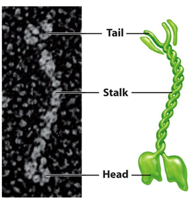
| Kinesin | |
| Kinesin Superfamily | G-Protein |
| Direction of Kinesin movement | From minus end to plus end |
| Structure of Kinesin 1- Chains | 2 light (70kDa) and 2 heavy (120kDa) |
| Design of Kinesin 1 - Cargo binding | Near C terminus |
| Design of Kinesin 1 - Stalk | Coiled coil structure, connecting cargo and microtubule binding sites |
| Design of Kinesin 1 - Microtubule binding sites | N terminus- 2 NTPase head motors |
| Design of Kinesin 1 - Neck Linkers | Connect NTPase heads to coiled coil stalk |
| Role of Neck linker | For power stroke |
| Role of coiled coil stalk | Connectivity and control |
| Sliding assay | Bind protein to glass, and see if it can move free microtubule |
| Rate of Kinesin movement | 800nm/second |
| Processive movement | Movement that can only occur by following a strict process |
| Monomeric heavy chain kinesin mutants | Slow and non-processive |
| Potential Models for movement of Kinesin | Inchworm and Hand Over Hand models |
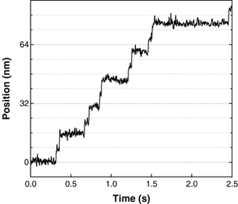
| Optical Trap | Use latex bead as cargo and use this method to measure it's movement |
| Inchworm Model | First head moves, second one catches up in 8nm steps |
| Hand Over Hand model | Each head moves 16nm- leading and trailing heads swap over |
| Determining Kinesin Movement Method | Label one head of Kinesin with a fluorophore and see how far it moves |
| Precision of Fluorophore Method | 1nm |
| Shows long lived, non moving stable state and 16nm steps | |
| Role of Switch's I & II | Detect presence of terminal (gamma) phosphate of ATP |
| Movement of Switch II | A spring loaded gate moving in/out in response to gamma phosphate |
| ATP Binds to sensor | Engaging it- phosphate release, releases sensor |
| ATP binding & sensor relationship | ATP binding engages sensor and vice versa |
| Relay Helix | Relays movement of Switch II around the protein |
| Conformational changes of ATP binding | Neck linker and tubular binding domain |
| 12 amino acid loop | Binds kinesin to tubulin |
Want to create your own Flashcards for free with GoConqr? Learn more.